'%3e%3cpath%20d='M706.453%20375.999C860.234%20375.999%20984.899%20291.829%20984.899%20188C984.899%2084.1703%20860.234%200%20706.453%200C552.671%200%20428.006%2084.1703%20428.006%20188C428.006%20291.829%20552.671%20375.999%20706.453%20375.999Z'%20fill='%23009B4F'/%3e%3cpath%20d='M278.447%20375.999C432.229%20375.999%20556.893%20291.829%20556.893%20188C556.893%2084.1703%20432.229%200%20278.447%200C124.665%200%200%2084.1703%200%20188C0%20291.829%20124.665%20375.999%20278.447%20375.999Z'%20fill='%23009B4F'/%3e%3cpath%20d='M141.484%20166.68C125.01%20162.158%20114.996%20158.927%20114.996%20150.529C114.996%20143.422%20122.102%20138.9%20133.085%20138.9C144.068%20138.9%20155.051%20143.099%20164.095%20150.529L164.418%20150.852L165.388%20129.855C156.666%20125.01%20147.944%20121.78%20133.731%20121.78C123.394%20121.78%20114.027%20124.364%20107.243%20129.209C99.8137%20134.378%2095.9374%20142.13%2095.9374%20151.175C95.9374%20168.941%20110.796%20176.694%20123.394%20179.924C140.192%20184.446%20151.821%20188.323%20151.821%20198.659C151.821%20207.381%20144.068%20212.872%20131.47%20212.872C118.872%20212.872%20106.274%20207.381%2096.5834%20198.013L96.2604%20197.69L94.3223%20219.979C104.336%20226.762%20116.934%20230.316%20130.501%20230.316C142.13%20230.316%20152.144%20227.408%20159.25%20221.917C167.003%20216.103%20171.202%20207.704%20171.202%20197.367C171.202%20181.862%20161.511%20171.848%20141.484%20166.68Z'%20fill='white'/%3e%3cpath%20d='M264.557%20188C264.557%20215.78%20246.467%20230.316%20225.471%20230.316C215.457%20230.316%20207.381%20226.762%20201.244%20221.271V253.896H182.186V146.976H201.244V156.02C207.704%20149.883%20216.103%20145.684%20226.763%20145.684C247.436%20145.36%20264.557%20160.22%20264.557%20188ZM245.498%20188C245.498%20172.494%20236.131%20162.158%20222.887%20162.158C214.811%20162.158%20207.058%20165.711%20201.567%20173.787V203.182C207.058%20210.288%20214.488%20213.841%20222.887%20213.841C236.131%20213.841%20245.498%20203.505%20245.498%20188Z'%20fill='white'/%3e%3cpath%20d='M352.095%20194.137H294.274C296.535%20207.058%20305.903%20213.841%20318.824%20213.841C329.807%20213.841%20337.882%20211.257%20346.604%20206.089L347.573%20222.886C339.82%20227.085%20330.13%20230.316%20317.532%20230.316C294.274%20230.316%20274.893%20215.78%20274.893%20188.323C274.893%20162.804%20294.274%20145.683%20317.209%20145.683C341.112%20145.683%20353.71%20160.22%20353.71%20181.216C353.71%20185.415%20353.064%20189.615%20352.095%20194.137ZM335.621%20178.632C335.621%20168.618%20329.161%20160.866%20317.532%20160.866C306.549%20160.866%20296.858%20168.295%20294.597%20180.893H335.298C335.621%20180.247%20335.621%20179.601%20335.621%20178.632Z'%20fill='white'/%3e%3cpath%20d='M443.189%20221.917L445.128%20203.505C452.88%20209.965%20463.54%20213.841%20474.846%20213.841C482.921%20213.841%20488.413%20210.611%20488.413%20204.797C488.413%20189.615%20444.482%20197.367%20444.482%20169.91C444.482%20153.436%20458.695%20145.36%20475.492%20145.36C487.444%20145.36%20496.488%20148.268%20503.272%20152.467L502.303%20169.587C494.227%20164.096%20484.213%20161.189%20475.492%20161.189C468.708%20161.189%20463.217%20163.773%20463.217%20168.941C463.217%20181.862%20507.471%20175.402%20507.471%20203.828C507.471%20221.271%20492.289%20229.993%20474.523%20229.993C461.602%20230.316%20450.942%20227.085%20443.189%20221.917Z'%20fill='white'/%3e%3cpath%20d='M590.488%20177.986V228.701H571.429V220.302C564.969%20226.439%20556.57%20229.993%20546.88%20229.993C528.467%20229.993%20517.484%20219.01%20517.484%20203.505C517.484%20187.03%20530.405%20177.017%20549.464%20177.017C556.57%20177.017%20563.677%20178.309%20571.752%20180.57V177.986C571.752%20165.388%20562.385%20159.897%20550.756%20159.897C542.034%20159.897%20532.02%20163.127%20524.914%20168.941L524.268%20152.467C530.405%20148.268%20541.711%20145.037%20554.309%20145.037C574.983%20145.36%20590.488%20155.374%20590.488%20177.986ZM571.429%20206.735V194.46C564.646%20192.199%20558.185%20190.907%20551.402%20190.907C541.711%20190.907%20535.574%20196.075%20535.574%20203.505C535.574%20210.934%20542.034%20215.78%20550.433%20215.78C558.508%20215.78%20564.969%20212.872%20571.429%20206.735Z'%20fill='white'/%3e%3cpath%20d='M592.104%20146.976H612.777L636.358%20198.659L659.616%20146.976H680.289L641.526%20228.701H630.866L592.104%20146.976Z'%20fill='white'/%3e%3cpath%20d='M754.907%20194.137H697.409C699.67%20207.058%20709.038%20213.841%20721.959%20213.841C732.941%20213.841%20741.017%20211.257%20749.739%20206.089L750.708%20222.886C742.955%20227.085%20733.264%20230.316%20720.667%20230.316C697.409%20230.316%20678.027%20215.78%20678.027%20188.323C678.027%20162.804%20697.409%20145.683%20720.343%20145.683C744.247%20145.683%20756.845%20160.22%20756.845%20181.216C756.522%20185.415%20756.199%20189.615%20754.907%20194.137ZM738.756%20178.632C738.756%20168.618%20732.295%20160.866%20720.667%20160.866C709.684%20160.866%20699.993%20168.295%20697.732%20180.893H738.433C738.433%20180.247%20738.756%20179.601%20738.756%20178.632Z'%20fill='white'/%3e%3cpath%20d='M826.618%20148.914L824.034%20166.357C819.511%20163.773%20814.989%20163.127%20810.467%20163.127C802.068%20163.127%20794.315%20167.326%20788.824%20176.371V228.701H769.766V146.976H788.824V157.958C794.315%20150.529%20802.068%20145.36%20812.728%20145.36C817.896%20145.36%20822.418%20146.653%20826.618%20148.914Z'%20fill='white'/%3e%3cpath%20d='M832.109%20221.917L834.048%20203.505C841.8%20209.965%20852.46%20213.841%20863.766%20213.841C871.841%20213.841%20877.333%20210.611%20877.333%20204.797C877.333%20189.615%20833.401%20197.367%20833.401%20169.91C833.401%20153.436%20847.615%20145.36%20864.412%20145.36C876.364%20145.36%20885.408%20148.268%20892.192%20152.467L891.223%20169.587C883.147%20164.096%20873.133%20161.189%20864.412%20161.189C857.628%20161.189%20852.137%20163.773%20852.137%20168.941C852.137%20181.862%20896.391%20175.402%20896.391%20203.828C896.391%20221.271%20881.209%20229.993%20863.443%20229.993C850.522%20230.316%20839.862%20227.085%20832.109%20221.917Z'%20fill='white'/%3e%3cpath%20d='M429.298%20206.412C422.838%20211.257%20416.054%20213.841%20408.302%20213.841C395.058%20213.841%20383.429%20204.474%20383.429%20188C383.429%20171.525%20395.058%20162.158%20408.302%20162.158C416.054%20162.158%20422.838%20164.742%20429.298%20169.264C430.268%20163.45%20431.237%20157.958%20432.852%20152.467C426.068%20148.591%20417.67%20145.683%20406.041%20145.683C382.783%20145.683%20363.725%20161.512%20363.725%20188C363.725%20214.487%20382.783%20230.316%20406.041%20230.316C417.67%20230.316%20426.068%20227.085%20432.852%20222.886C431.237%20217.395%20429.944%20211.903%20429.298%20206.412Z'%20fill='white'/%3e%3c/g%3e%3cdefs%3e%3cclipPath%20id='clip0_1478_9029'%3e%3crect%20width='984.9'%20height='375.999'%20fill='white'/%3e%3c/clipPath%3e%3c/defs%3e%3c/svg%3e)
What is the eye?
The human eye is one of the most sophisticated organs in your body, with multiple parts working together to capture light, focus it, and convert it into electrical signals your brain interprets as vision. Your eye is like a tiny camera, and every part plays a role in helping you see.
How does the eye work?
To understand how the eye works, it helps to follow how light travels through your eye.
Cornea
This is a see‑through, dome‑shaped front layer that covers the iris, pupil, and anterior chamber. It provides about 70% of your eye's focusing power by refracting (bending) light as it enters. Irregularities in the cornea's shape can cause astigmatism, where light doesn't focus properly on the retina.
Iris and pupil
The iris is the coloured part of your eye containing muscles that control the pupil size. The pupil is the dark opening that allows light to enter. In bright conditions, the pupil constricts (gets narrower); in dim light, it dilates (gets wider) to let more light in.
Lens
Located behind the iris, the lens provides 30% of your eye's focusing power and can change shape for near and distance vision. As we age, the lens loses flexibility, leading to presbyopia, where near vision becomes difficult.
Retina
The light‑sensitive tissue lining the back of your eye that converts light into electrical signals. The retina contains photoreceptors (rods and cones) that detect light and colour, sending signals via the optic nerve to your brain.
Aqueous Humour
This is the clear fluid filling the front chambers of your eye. It maintains eye pressure, provides nutrients to tissues without blood vessels, and removes waste. Problems with drainage can lead to glaucoma.
Vitreous Humour
This gel‑like substance fills the large cavity behind the lens. It helps maintain the eye's shape and allows light to pass through to the retina. Changes with age can cause floaters and flashes.
Do these symptoms sound familiar?
If you have experienced any of the symptoms mentioned above it's worth booking an appointment with one of our optometrists.
What causes eye problems?
Problems with eye anatomy can affect different parts of your vision system. Issues with the cornea or lens can cause focusing problems, while retinal conditions can affect how light is processed. Understanding the anatomy of eye structures helps explain why conditions develop and how they're treated.
How are eye problems treated?
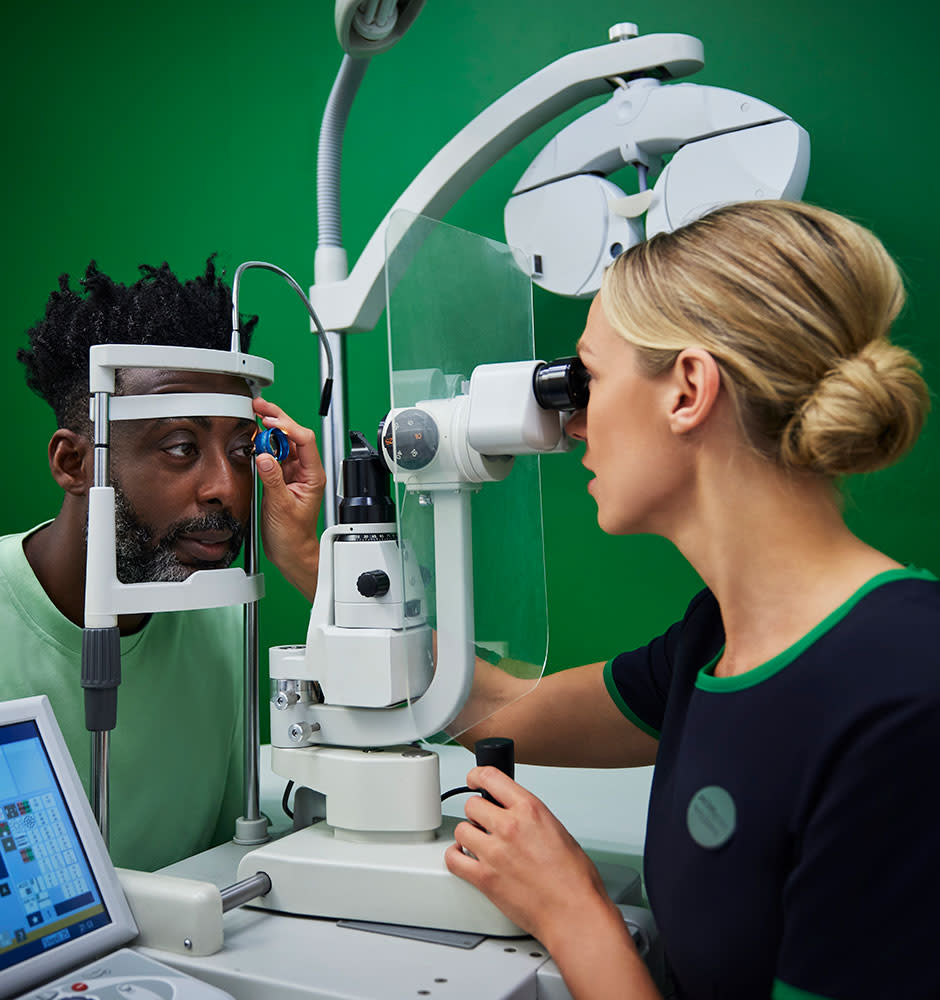
An optometrist can diagnose eye problems by examining all parts of your eye anatomy. They use specialised equipment to look at the front and back of your eye, test your vision, and check how well your eye muscles work together.
Regular eye tests help detect problems early when treatment is most effective.
Patient history: Your optometrist will ask about your symptoms, any previous eye problems, and your general health to understand your eye health.
Vision testing: Comprehensive tests check how well each part of your visual system works, including tests for refractive errors and colour vision.
Eye examination: Using specialised equipment to examine the cornea, lens, retina, and optic nerve for signs of disease or damage.
Can eye problems be prevented?
While not all eye problems can be prevented, you can take steps to protect your eye anatomy and reduce your risk. This includes wearing sunglasses to protect from UV damage, eating a healthy diet rich in vitamins, not smoking, and having regular eye tests to detect problems early.
Eye anatomy FAQs:
The main parts include the cornea (clear front layer), iris (coloured part), pupil (opening), lens (focusing element), retina (light‑sensitive back layer), macula (central vision area), optic nerve (connection to the brain), and supporting structures like muscles and fluids.
The iris is found behind the cornea and in front of the lens. It's the coloured part that gives you your distinctive eye colour and controls the pupil size to regulate light entry.
The aqueous humour is a clear fluid that fills the front chambers of your eye. It maintains eye pressure, provides nutrients to tissues without blood vessels, and removes waste products.
Six extraocular muscles control eye movement: four rectus muscles move the eye up, down, inward, and outward, while two oblique muscles provide rotational movements. They must work in perfect coordination.
The cornea is the fixed front layer providing 70% of focusing power, while the lens is behind the iris, providing 30% and can change shape for near and distance vision.
The macula is the small, central area of the retina responsible for detailed central vision, allowing you to read, recognise faces, and see fine detail.
The optic nerve carries visual information from the retina to your brain, where it’s processed into the images you see.
The iris muscles contract or relax to make the pupil smaller in bright light and larger in dim light, helping control how much light enters the eye.
Written by our experts for peace of mind

Ross Campbell
Related conditions
Not sure if this is what you’re experiencing? Explore some related conditions:
Blurry Vision
Blurry vision is not an eye condition itself, it’s usually a symptom of an underlying condition or a sign that your glasses or contact lenses may need updating.
Find out moreSquint
A squint, also known as strabismus, occurs when a person’s eyes point in different directions.
Find out moreAstigmatism
This is simply an imperfection in the shape of your eye that affects how you see.
Find out more
Find out more about eye care services
All eye conditions
Read more information about all eye conditions.
View all eye conditions
Book an eye test
Book an appointment at your local Specsavers store.
Book an eye test
All about eye tests
Everything you need to know about getting your eyes tested.
Find out more
Rehman, I. (2023) Anatomy, Head and Neck, Eye. In: StatPearls. Treasure Island (FL): StatPearls Publishing. Available at: Available at: https://www.ncbi.nlm.nih.gov/books/NBK482428/
(Accessed: 17 August 2025)Neuroscience Anatomy of the Eye. NCBI Bookshelf. Available at: Available at: https://www.ncbi.nlm.nih.gov/books/NBK11120/
(Accessed: 17 August 2025)Kolb, H. (2007) Gross Anatomy of the Eye. In: Webvision: The Organization of the Retina and Visual System. University of Utah. Available at: Available at: https://www.ncbi.nlm.nih.gov/books/NBK11534/
(Accessed: 17 August 2025)Kels, B.D. (2015) ‘Human ocular anatomy’, Clinical Anatomy, 28(1), pp. 71–99. Available at: Available at: https://pubmed.ncbi.nlm.nih.gov/25704934/
(Accessed: 17 August 2025)Willoughby, C.E. (2010) ‘Anatomy and physiology of the human eye: effects on ocular physiology’, Clinical & Experimental Ophthalmology, 38(2), pp. 93–101. Available at: Available at: https://onlinelibrary.wiley.com/doi/10.1111/j.1442‑9071.2010.02363.x
(Accessed: 17 August 2025)Ludwig, P.E. (2024) Physiology, Eye. In: StatPearls. Treasure Island (FL): StatPearls Publishing. Available at: Available at: https://pubmed.ncbi.nlm.nih.gov/29262001/
(Accessed: 17 August 2025)Kenhub ‘Eye anatomy: Muscles, arteries, nerves and lacrimal gland’. Kenhub. Available at: Available at: https://www.kenhub.com/en/library/anatomy/eye‑anatomy
(Accessed: 17 August 2025)American Academy of Ophthalmology (2023) ‘Eye Anatomy: Parts of the Eye and How We See’. AAO. Available at: Available at: https://www.aao.org/eye‑health/anatomy/parts‑of‑eye
(Accessed: 17 August 2025)Cleveland Clinic (2021) ‘Eyes: How They Work, Anatomy & Common Conditions’. Cleveland Clinic. Available at: Available at: https://my.clevelandclinic.org/health/body/21823‑eyes
(Accessed: 17 August 2025)